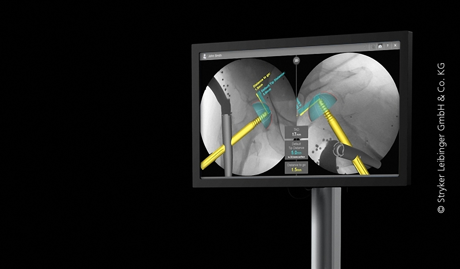
Stryker Corporation is one of the largest manufacturers of medical devices in the world and one of the leading companies in the area of orthopedic products. A computer assisted surgery system for minimally invasive treatment of hip fractures developed by Stryker Navigation in Freiburg celebrated its market launch at the end of 2011 after a four-year R&D phase, in which ITWM provided key support in the area of parameter identification.
A hip fracture is treated by implanting a rod, known as a nail, into the shaft of the femur bone and securing it in place with a surgical screw. To achieve the maximum biomechanical stability, it is important that the screw penetrate as far as possible into the ball portion of the hip joint. On the other hand, under no circumstance should the tip of the screw be allowed to penetrate the periosteum, so as not to restrict the mobility of the ball joint. Because this is a minimally invasive surgery, the operator is not able to see the relative positions of the screw and bone with his own eyes, but rather must rely on imaging methods.
3D-Reconstruction Predicts the Penetration Point of the Screw
Over the course of the past years, Stryker Navigation has developed a pioneering solution, one that both enables more precision work and requires only minor changes to the established procedures. This is accomplished using two X-ray images that can be produced using the common C-arm. In the process, a reference body with small beads that stand out well in the 2D X-ray images is attached to the nail, which also serves as the guide for the screw.
This enables a 3D reconstruction of the anticipated axis of the screw. By exploiting the ball shape of the femural head, it is possible to extract its contour, to reconstruct its spatial position, and to predict the penetration point of the screw.

