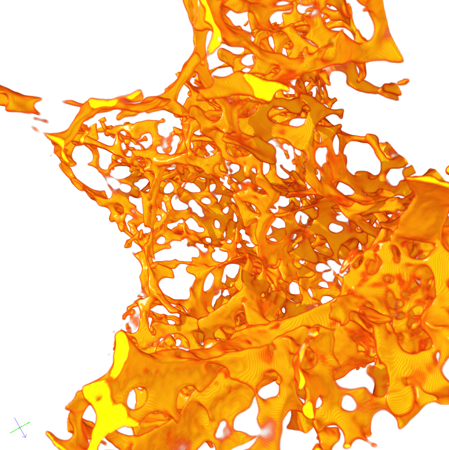
Capillary vessels are the smallest offshoots of the blood vessel system. They pervade the whole body, in particular all organs.
Here we mainly study capillary vessels in lung and liver. From their layout we can conclude in what condition the tissue and the actual organ are.
3D-Analysis of the Vessels
So far the examination of these extremely small and dense vessels was limited to visual analysis of 2D microscope photographs. However, the analysis in 3D provides more exact information, especially concerning the collocation. Furthermore, the high resolution (400 – 700 nm) yields a detailed representation of the highly branched structure. This paves the way for extensive findings in medical research.
First of all, the vessels are filled with a special plastic and the surrounding tissue is removed using acid. What remains is recorded with high resolution Synchroton Tomography.
Adjust Segmentation Processes
The graphic material now has to be segmented to capture the vessel structure exactly. It is necessary to adjust already existing segmentation processes. A quantitative analysis of the images yields the exact volume and surface area of the vessels and the topology of the vessel structure. Using the Euler number one can find small holes in the capillary vessels. These indicate how big the compensatory lung growth is, a process where lung can regrow after up to 40% is removed. As this is a process in mice lungs, findings could help improving therapy methods for diseases of the human lung, e.g. cancer.
Our main task is to develop algorithms to record and describe the structure of the vessel system.

